Supported by Dr Robbie Goodrum DO from the UK
The Radiology program is a 2 x 5 day courses. Studying from 9-5 pm daily.
You will need to study several radiology books before this program starts as a pre study.
Contact us regarding books to study
Tutors are medical radiologist Doctors.
1.Congenital Anomalies and Normal Variants,
Occipitocervical fusion
Paramastoid process
Agenesis/hypoplasia
Posterior ponticle
Os odontoideum
Ossiculum terminale
Congenital aplasia and dysplasia of the densI
Intercalary bone
Nuchal bone
Stylohyoid ligament ossification
Block vertebrae
Klippel-Feil syndrome
Sprengel’s deformity
Spina bifida
Cervical ribs
Costal cartilage calcification
Hahn’s fissures
Knife-clasp deformity
Transitional segments
Butterfly vertebra
Hemivertebra
Asymmetric facets
Absent pedicle
Limbus bone
Nuclear impression
Schmorl’s node
Os acetabulum
Fabella
Bipartite, tripartite and multipartite patella
Madelung’s deformity
Fibrous dysplasia
Complete occipitalization.
2.Arthritides
Degenerative arthropathies
Inflammatory arthropathies

Ankylosing spondylitis
3.Tumor
Benign tumor of bone
Malignant tumor of bone
Diffuse osteoporosis of multiple myeloma pathologic vertebral body fracture
4.Blood(Vascular)
Sickle cell anemia
Thalassemia (Cooley’s anemia)
Avascular necrosis (ischemic necrosis)
Osteochondroses
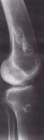
Distal femur and proximal tibia shows focal medullary avascular necrosis.
5.Infection
Suppurative osteomyelitis
Infectious arthropathies
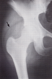
Infection shows the displacement of the gluteus medius fat plane
6.Trauma
Types of fractures
Orientation
Spatial relationships of fractures
Traumatic joint disruptions
Fracture repair
Complications of fracture
Interpretation of radiographs in the evaluation of fracture healing
Fractures of the skull
Spinal trauma
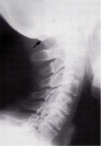
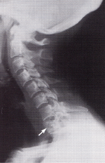
Cervical spine
Cervical spine fractures
Thoracic and lumbopelvic fractures
Sacral injuries
Coccygeal injuries
Fracture and dislocations of the appendicular skeleton
Spondylolisthesis
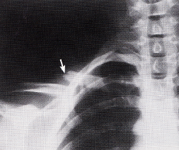
A misaligned fracture of the clavicle
7.Endocrine and Metabolic Bone Disease
Osteoporosis
Osteomalacia
Rickets
Hyperparathyroidism
Cushing’s disease and steroid-induced osteonecrosis
Paget’s disease
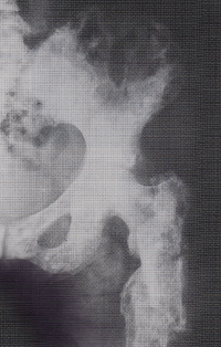
Paget’s disease.
8.Soft Tissue
Concretions
Conduit wall calcifications
Cyst wall calcifications
Solid mass calcifications
Location of abdominal calcifications
Calcification that may cross the midline
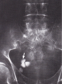
radiopaque urinary vesicular calculi
9.Scoliosis
Structural and nonstructural scolioses
Complications
X-ray assessment
Mensuration
Therapy selection
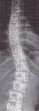
Observe the scoliosis & multiple vertebral body rotation in the left thoracolumbar convexity
Fee $1500USD per 5 day course
$ 2,800 for both courses.
Note this Radiology course is part of a 3 year part time Degree in Osteopathy. The 3 year Doctor of osteopathy degree is organized by Goodrum Seminars in association with the National University of Medical Science & the Indian Osteopathic institute.
This module can be taken as a CPD post graduate Seminar.
Course dates Booking form testimonials